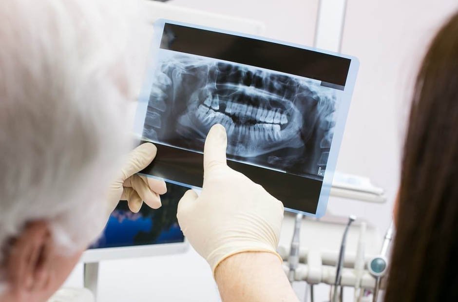
As parents, we are constantly worried about our kids’ welfare, particularly regarding their health. Pediatric dental care is essential to preserve a child’s general health, and X-rays are great resources for identifying and treating various oral health problems. However, it is understandable that the word “radiation” can cause apprehension.
Dental X-rays are crucial to thorough dental exams because they provide dentists access to structures otherwise hidden from vision. They aid in finding cavities, tracking tooth growth, evaluating the roots, and spotting future orthodontic issues. These diagnostic images provide insightful information that helps with early detection and treatment, thereby avoiding more serious oral health issues in the future.
This article will examine the world of pediatric dental X-rays, look at the several X-ray types used in dentistry, and—most importantly—discuss the precautions that may be taken to reduce radiation exposure and protect your child.
What are Dental X-Rays?
Dental X-rays, also known as radiographs, are a vital diagnostic tool that allows dentists to view parts of the mouth not visible to the naked eye. These images can reveal decay between teeth, problems in the root areas, and issues pertaining to bone density. Especially for children, dental X-rays help monitor growth and development, ensuring teeth come in properly. They also assist in determining if all primary teeth have fallen out to make room for permanent teeth.
When it comes to children’s dental health, expertise is paramount. Junior Smiles of Stafford, a leading pediatric dentist in Woodbridge, VA, employs the latest X-ray technologies to ensure minimal exposure and maximum insight, safeguarding your child’s health while delivering comprehensive dental care.
Different Types of Dental X-Rays
Bitewing X-Rays
These images focus on the upper and lower back teeth. Bitewings are useful for detecting decay between teeth and assessing bone health.
Periapical X-Rays
Providing a detailed view of a single tooth, periapical X-rays help identify issues within the tooth root and surrounding bone.
Panoramic X-Rays
These broad images capture the entire mouth, including teeth, jaws, and sinuses, making them helpful for orthodontic evaluations and detecting impacted teeth.
Radiation Risks in Pediatric Dental X-Rays
Radiation exposure, a critical concern in medical imaging, is quantified in units known as millisieverts (mSv). Compared to other medical imaging techniques, dental X-rays typically use lower doses of radiation. Still, it’s important to remember that children are more vulnerable to radiation’s side effects than adults.
The safety and the worthwhile advantages of early diagnosis and treatment must be balanced, notwithstanding the possible radiation risks. Dental X-rays are essential for detecting oral health concerns early on, allowing for prompt treatment and averting the emergence of more severe disorders. Early detection can result in more efficient and minimally intrusive treatments, improving your child’s oral health.
Dental experts adhere to strict criteria and practices to safely perform dental X-rays. These recommendations are made to minimize radiation exposure while obtaining precise and trustworthy diagnostic images. The safety of the child is given top priority by dental staff during the X-ray procedure because it is always the primary concern.
Best Practices to Minimize Radiation Exposure
Use of Lead Aprons and Thyroid Collars
When taking X-rays, lead aprons and thyroid collars should always be used in pediatric dental offices. Thyroid collars protect the thyroid gland from exposure, while lead aprons shield the child’s body, reproductive organs, and other essential areas from unneeded radiation.
Selective Use of X-Rays
Dentists must exercise discretion in ordering X-rays. A decision should be made based on the child’s dental history, age, risk factors, and present oral health condition. The dentist should request the child’s records if recent X-rays were taken elsewhere on the youngster to prevent needless duplication.
Digital Radiography
Modern dental practices use digital radiography, dramatically lowering radiation exposure. Digital systems require atleast 90% less radiation, resulting in safer imaging for young patients.
Collimation and Rectangular Beam
Collimation limits the X-ray beam to the region of interest, minimizing exposure to nearby tissues. By restricting the X-ray beam to the shape of the dental receptor and removing extraneous scatter radiation, rectangular collimation significantly decreases radiation.
Fast Film or Sensor Speed
The speed of dental film or sensor used during X-rays impacts the amount of radiation needed to produce an image. Faster film or sensors require less radiation, minimizing exposure time for the child.
Focused X-Rays
Precise aiming of the X-ray beam ensures that only the necessary areas are exposed, minimizing radiation scatter.
Proper Training and Certification
Dental professionals must undergo extensive training and certification. Properly trained staff ensures that X-rays are taken accurately and efficiently, limiting the need for retakes and additional radiation exposure.
Guidelines for Parents
Share Relevant Medical Information
Parents should inform the dental team of any known allergies or medical conditions that might affect the child’s ability to undergo X-rays safely.
Ask Questions
Feel free to ask the dentist about the type of X-rays recommended, why they are necessary, and how they plan to minimize radiation exposure.
Final Thoughts
Pediatric dental X-rays are essential for preserving your child’s oral health. As parents, you can ensure that your child receives the greatest dental treatment while maintaining their general well-being by participating in the procedure and being informed of the safety precautions implemented.
Pediatric dental X-rays can be made safer by working with an experienced dental team and informed parents to improve our children’s long-term health.


